
*Country restrictions apply
This simple to use microscopy-based test contains all of its staining agents within a single tube, thereby making testing faster and easier.
Clinicians on the front line of the malaria battle need fast results. The QBC® Malaria Test is a highly respected, sensitive and specific laboratory-based test for any technician capable of performing traditional thin or thick films.
The test itself can be prepared in just six to seven minutes, as it requires only to be filled with capillary or venous blood then centrifuged in the QBC Centrifuge for five minutes. The test can then be analysed under a fluorescent microscope (we recommend our QBC ParaLensTM Advance Fluorescent Microscope System attached to your existing light microscope).
Benefits Over Conventional Malaria Diagnostics
- Increased sensitivity
- Increased specificity
- Reduced training times
- Time-saving
- Simple set up and review
- Respected, proven technology
- Ideal for use in the field
- Safe fluorescence - No mercury when using the QBC ParaLens Advance
QBC Malaria Tube
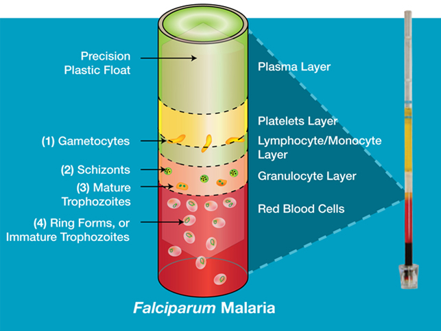
The QBC Malaria Tube is internally coated with all the necessary reagents and anticoagulants to perform a malaria test in minutes. The QBC Malaria Tube contains a fluorochrome called acridine orange and anticoagulants - when a capillary or venous blood sample is added, a precision-engineered plastic float is inserted into the QBC Malaria Tube. When the QBC Malaria Tube is centrifuged, the blood becomes concentrated and the precision float sits level with the Buffy Coat layer. The blood components and malaria parasites separate based on density and concentrate in distinct layers. Parasites and leucocyte nuclei fluoresce a yellowish-green. It is in the Buffy Coat layer where the malaria parasites will be visible as shown below:
Malaria Parasite Identification
The QBC Malaria Tube can be used to detect malaria parasites:
- The QBC Malaria Tube is placed in the QBC ParaViewer, allowing the tube to be rotated during microscopy so that the whole diameter of the tube may be examined - not just a one-dimensional view
- 24 “fields” can be examined by rotating the tube during examination
- Once in position, the tube may be examined for malaria parasites at the upper end of the float and for other parasites by moving down to the other end of the tube
- Red blood cells (RBC's) containing malaria parasites are less dense than non-infected RBC’s
- In light malaria infections, following centrifugation, the parasitised RBC’s form the upper layer (the “brown” layer) of RBC’s - most of the malaria parasites will be found here. In heavy malaria infections, parasites may be found anywhere in the tube
- Due to its density, the float, inserted into QBC Tube before centrifugation, will settle between the upper RBC layer and the upper plasma area. The float creates a monolayer enabling fluorescent parasites to be easily seen
The QBC Malaria Tube is easy to use and has a significantly reduced training time compared to thick and thin films, taking just five days to become proficient in identifying parasites.
Other Blood Parasite Identification
The QBC Malaria Tube can also be used to detect other blood-borne parasites:
- The QBC Malaria Tube is the gold standard test in diagnosis of T.b.gambiense in blood - Trypanosomes are killed by acridine orange so tubes need to be examined immediately after centrifugation to see motile trypomastigotes
- Microfilariae and Borrelia may be seen anywhere in QBC Tube following centrifugation
The QBC Malaria Tube can detect various different parasites - please view our Scientific Papers page for further details.
Using the QBC Malaria Tube under an LED fluorescent microscope (such as the QBC ParaLens Advance) identification of the following parasites is possible:
| 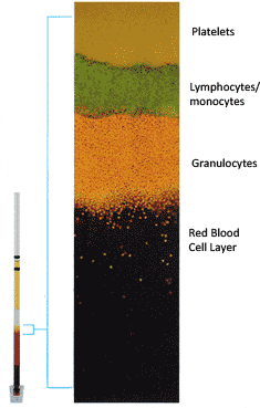 |
The float occupies the area midpoint between red cells and plasma. The amber layer surrounding the float is the Buffy Coat. Seen enlarged above, the three separate layers of the Buffy Coat are comprised of orange-yellow fluorescing platelets (top), green lymphocytes/monocytes (middle) and yellow granulocytes (bottom). Below the granulocytes is the red blood cell layer (some overlap of RBC's and granulocytes occurs). Red cells containing Plasmodium parasites concentrate at the top of this layer and appear as multiple "pinpoints" of fluorescence. A negative specimen shows no fluorescing points in this area.
Method
55-110µl of blood is taken in a QBC Malaria Tube which is internally coated with acridine orange and centrifuged for five minutes. When viewed under a fluorescent microscope, the malaria parasites are seen concentrated, shining in the background of dark Red Blood Cells.
Sensitivity
The most sensitive test available for the diagnosis of malaria due to:
- The high volume of blood
- The concentration of the parasites by centrifugation
- The fluorescent staining of parasites
The QBC Malaria Test is 5.5 to 7% more sensitive than Giemsa thick films. It can detect as little of one parasite per µl of blood and establish diagnosis earlier than thick film in 47% of low parasitemia (<10 parasites per µl) cases. Up to 18% more positives are reported by QBC than the conventional thick film method.
Diagnosis
All Plasmodium species are identified and quantified in the QBC Malaria Tube using the QBC ParaLens Advance system (or traditional microscope). Species differentiation is possible in the QBC Malaria Test, enabling the physician to administer the right type of medication. Because of the high sensitivity of the QBC test, it is possible to detect malaria parasites very early, avoiding a long recovery and loss of working days for the patient. A QBC test is highly specific and a negative result will enable the patient to avoid a multi-drug therapy.
QBC ParaLens Advance - Low-Cost Alternative to a Fluorescent Microscope
The QBC Malaria Tubes are for use with a fluorescent microscope. The QBC ParaLens Advance can be attached to a standard light microscope to create an LED fluorescent microscope; a low cost and more portable alternative to a traditional fluorescent microscope. The QBC ParaLens Advance utilises LED fluorescence microscopy.
Ordering Information
| Product Code | Product Description |
| 253037 | Malaria Tubes (100) |
| 253005 | Malaria Tubes (2000) |
If you would like more information please call +44 (0) 1204 460446 or email sales@woodleylabdiagnostics.com